Participant Preparation
Preparation procedures can be done by both researchers and operators. The intention is for researchers to take the lead in this process using operator's help only when required. If you are not sure how to do the preparation, read these guidelines, and ask operators for further coaching. If you actively participate in the preparation, you can use your recording times much more efficiently: e.g., you can already prepare the next subject, when the operator is finishing the previous recording.
If you are also going to record EyeTracking data, you will have to make sure the participant isn't wearing any mascara. Using the eye tracker with the MEG compatible glasses is very difficult and is not recommended. Contact lenses are no problem.
Checks and wires
Subjects need to be free of magnetic materials, like coins, watches and other metal items. Underwired bras can be a problem, and even dyes in clothing or hairdye can cause interference.
It is recommended that researchers take their participants through a MEG Subject Screening Form, which will prompt them to remove all metal. This form can be downloaded from here: MEG SOPs
To make sure that a subject is non-magnetic, it is good practice to put them in the MEG machine before any other preparation, and to check the signal for artefacts.
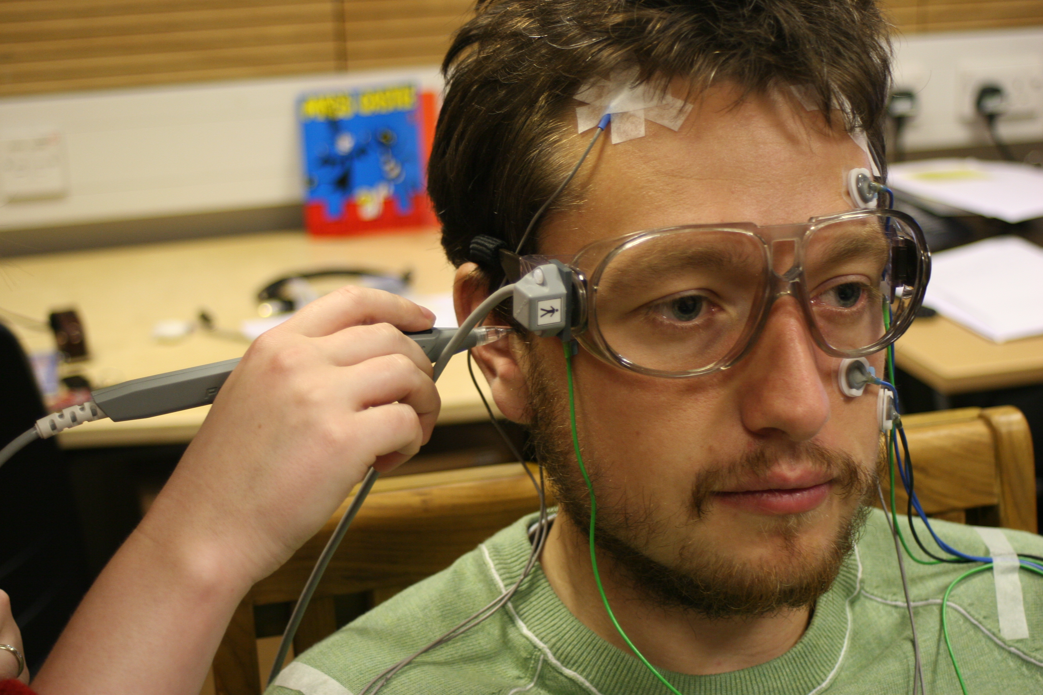
To track head movements volunteers need to have 5 small HPI (Head Position Indicator) coils attached to their head: 2 on the forehead, 2 behind the ears and one on the crown. When using an EEG cap, the HPI coils are attached to the cap. In addition we normally attach 5 electrodes to the face as well to measure eye movements and blinks.
The location of the coils needs to be recorded with a Polhemus 3D digitiser. We also measure 3 landmarks and a number of additional points on the head to indicate headshape and enable easier matching to an MRI structural scan. The digitiser has a pen-like sensor that will record the location of its tip when the button is pressed. Please read our MEG Digitisation Procedure for more information.
In total, subject preparation should take about 20-30 minutes when no EEG is being recorded, and about an hour when a full EEG cap is being used.
The glasses in the picture have an additional sensor attached to them, to correct for movements of the head. It is essential that these glasses remain stable throughout the entire digitisation process.The cable leading to the glasses should be fixated to the chair. During digitisation, conversation with the participant should be kept to a minimum. Mobile phones and other metal objects should be removed from the participant as well as the experimenter during digitisation.
We routinely use the right and left preauricular (RPA, LPA) points and the nasion as anatomical landmarks. In order to make sure that data sets are comparable across participants (both within and across studies), we encourage everyone to use the following locations as accurately as possible (the photos are also on display in the MEG preparation room):
LPA: RPA:
Nasion:
Seating the subject in the MEG
This is always performed by a qualified operator.
It is important that subjects are as high in the helmet as possible. Most people will relax during the experiment and often end up in a slightly lower position than initially. It is good practice to allow for a bit of time to seat the subject properly, perhaps try to add or remove one or more pillows and make sure they are as comfortable as possible.
With very small volunteers they might end up being too much to the back, relative to the helmet. Smaller volunteers can then end up with their head uncomfortably pushed forward by the helmet. We have available a booster seat to help with positioning smaller participants effectively and the ability to move the whole chair forward slightly by using the levers on the side of the MEG machine.
With volunteers that have mobility problems we have to be extra careful.The best thing is to pull the chair out from under the dewar completely and to raise it up to the level of the participants knees or higher when the subject is getting in or out of it. It is essential that the brake is applied before asking your participant to sit and to make sure that the legrests are down.

