Checking for artefacts
Spin history and motion
Description
During each acquisition of a slice spins are excited, and then gradually relax back into line with the magnetic field with a rate determined by the T1 constant. In a typical FMRI design, the TR is not much larger than the T1, and so the spins will not relax back completely by the time the next acquisition arrives. This is usually dealt with by having a number of "dummy scans" at the start of each acquisition, to allow time for equilibrium in the spin excitation. If there is a sudden motion in the subject half way through a scan, a particular slice may correspond to a different part of brain than it did last time. It will be in a different degree of excitation, and the signal intensity will be different.
When it will be worst
When you have large subject motion with an interleaved acquisition.
What to look for
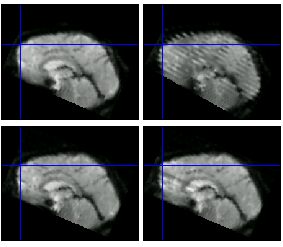
In the worst case scenario, if using an interleaved acquisition and the subject moves up by exactly one voxel, what were the odd slices become the even ones, and instead of all slices being acquired previously 2 seconds before, they alternate between 1 s and 3 s. Striping in the z direction will result.
|
Top row- interleaved sequence; bottom row- descending sequence; left column without movement, right column with movement. The spin history effect leads to striping throughout the image with the interleaved sequence, but only a single stretched part with the descending sequence |
You can often see this artefact as a peak in the variance visible in the top tsdiffana graph. For a more sensitive assessment, Daniel Mitchell at the CBU has written a tool for helping automatically find this striping.
RF spiking
Description
The MR machine is a sensitive radio receiver. Any spiking source of RF interference (e.g., static electricity) can lead to image artefacts. RF artefacts are rare, but you should be alert for them.
What to look for
Regular striping. Here is an extreme example from an MRI website - the bottom image is a simulated slice from a phantom. http://www.revisemri.com/tools/kspace/k_bigrfperiph.htm
Last updated by Rhodri Cusack Mar 2006
